Rohan prepping for surgery
Hello Chin lab!
Annie and Dan’s paper featured on the cover of Cell Reports
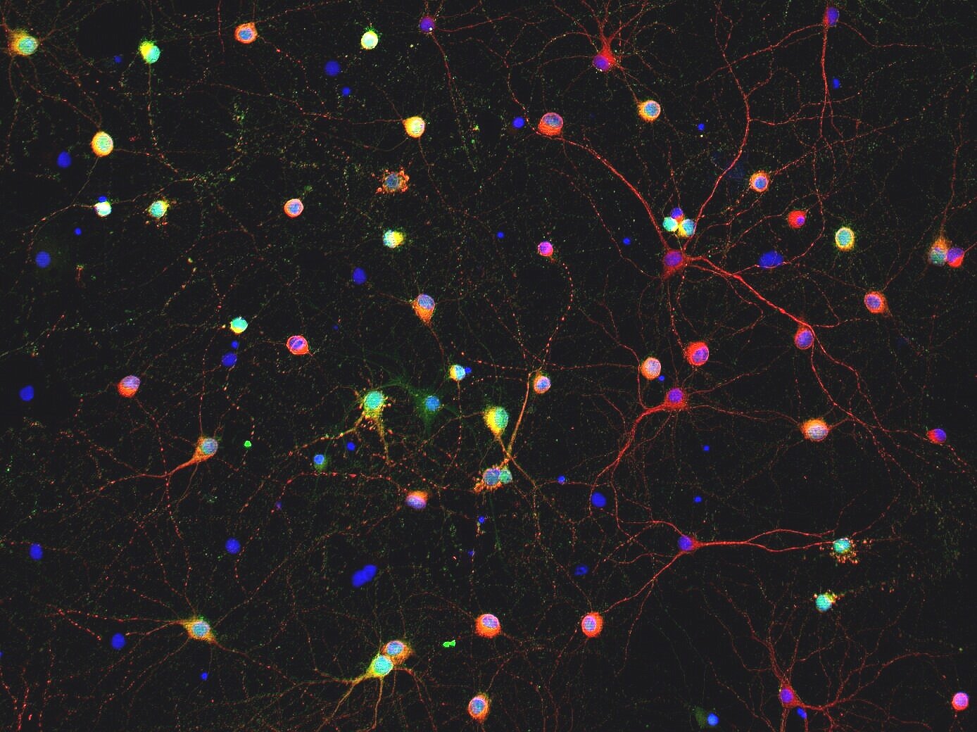
Primary hippocampal neurons stained for ∆FosB and MAP2 (Jason You, MD/PhD)
Double-labeling of ∆FosB and calbindin in the dentate gyrus (Jason You, MD/PhD)
Gabe at the bench
Annie in the microscope room
Immunophenotyping uses morphology and gene marker expression to assess cell types (Annie Fu and Dan Iascone)
Rohan’s paper featured on the cover of the Nov 3 issue of Science Translational Medicine
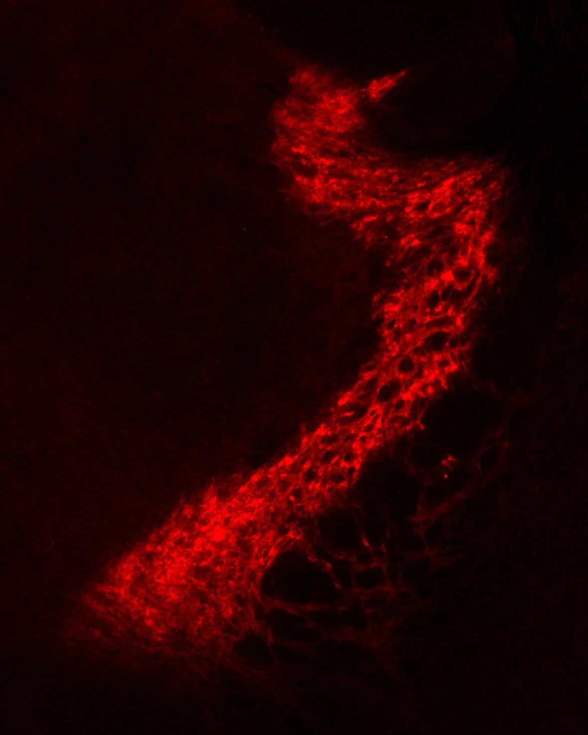
The thalamic reticular nucleus, expressing mCherry in TRN neurons. (Rohan Jagirdar, PhD)
Rendering of the TRN and EEG traces from the same animal (Rohan Jagirdar, PhD)
Slow ocean waves representing the “slow waves” of neuronal activity that make up slow wave sleep. (Jeannie Chin, PhD)
The thalamic reticular nucleus, expressing mCherry in TRN neurons, with the rest of the brain stained with DAPI. (Rohan Jagirdar, PhD)
Hello Chin lab!! … at the 33rd Rush and Helen Record Neuroscience Forum in Galveston (San Luis Hotel & Conference Center)


